Mr Brown is seen two months later and feels his vision is slightly worse.
The ophthalmologist does an OCT scan. OCT scans are used widely in ophthalmology to look at retinal disease. These are cross sectional scans which show the layers of the retina.
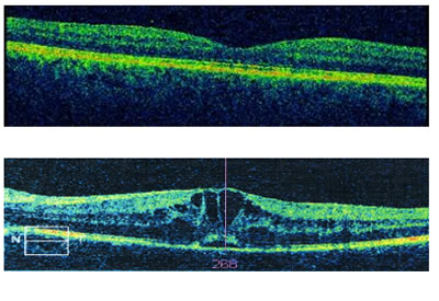
The picture at the bottom shows black cystic spaces (abnormal). Compare this to the normal scan at the top demonstrating the normal “dip” at the fovea.
This demonstrates macular oedema which can compromise vision further.