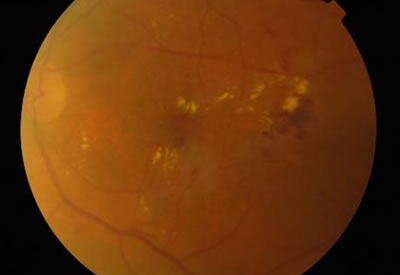
Diabetic Retinopathy
The fundus would show microaneurysms, blot haemorhages as well as hard exudates at the macula. It might also show cotton wool spots. The major vessels would not be tortuous and the disc margins shouldn’t be blurred.
Glossary
Cotton Wool Spot – Leakage of axonal material due to ischaemia
Microaneurysms – Small blood vessel dilatations due to endothelial damage in diabetes
Blot Haemorrhage – leakage of blood from dysfunctional blood vessels
Hard Exudate – Leakage of lipid material from dysfunctional blood vessels
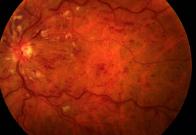
Central Retinal Vein Occlusion
Key features:
1. Swollen disc
2. Tortuous vessels
3. Widespread “flame-shaped” haemorrhages
There may also be cotton wool spots as shown.
Think Stormy Sunset
Retinal arteries and veins travel together, if the artery is atherosclerotic it can compress the vein and precipitate venous thrombosis.
Branch Retinal Vein Occlusion
The fundus would show flame haemorrhages usually confined to one quadrant only as only one branch is affected.

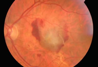
Central Retinal Artery Occlusion
Central retinal artery occlusions present with profound visual loss. There is likely to be a relative afferent pupillary defect and the fundus shows the characteristic “cherry red spot” – arrowed. The rest of the retina is pale.
The cherry red spot represents a window lnto the choroidal circulation which has its own blood supply, and therefore unaffected, at the thinnest part of the retina, the fovea.
It usually arises as a result of an embolus e.g. from carotid artery atheroma.
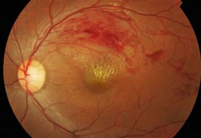
Wet AMD
Although Wet AMD also presents quickly and in the older age group, fundus findings of haemorrhage (red blood in picture), exudate (creamy area in picture) and fluid are confined to the macula only.
There is no disc swelling and vessels are not tortuous.